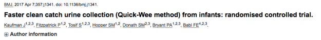
Where can I find this paper?
https://www.ncbi.nlm.nih.gov/pubmed/28389435
Please note this paper is OPEN ACCESS. You are strongly advised to read the original paper before reading any further.
What is this paper about (what is the research question)?
Does suprapubic cutaneous stimulation with cold fluid-soaked gauze (the “Quick-Wee” method) reduce the amount of time spent waiting for clean catch urine?
Summary of the Paper
Design: single centre, randomised, prospective non-blinded trial
Objective: to evaluate the efficacy of the Quick-Wee method
Outcome of interest: voiding of urine within five minutes (binary outcome)
Intervention: genital cleaning for 10 seconds with sterile water at room temperature, followed by continued rubbing of the suprapubic area in a circular pattern with gauze (soaked in cold saline) held by forceps
Reference standard: genital cleaning for 10 seconds with sterile water at room temperature (standard practice)
Participants: patients presenting to an Australian paediatric emergency department between September 2015-April 2016
Inclusions: pre-continent infants aged 1-12 months in whom clean catch urine sample was required
- Exclusions: neonates (defined as <1 month of age); infants with anatomical or neurological abnormalities affecting voiding of urine or sensation; those patients with need for an immediate sample by invasive method
Results: 354 subjects were recruited of whom 344 participated in the analysis; 175 in the control group and 179 in the intervention group (5 patients were excluded from each group after randomisation, giving 170 in the control group and 174 in the intervention group).
54/174 (31%) of patients voided within five minutes in the Quick-Wee group
20/170 (12%) of patients voided within five minutes in the control group
The difference in proportions was 19% (95% confidence interval for difference 11-28%).
This gave an NNT of 4.7 to successfully catch one additional sample within five minutes (95% confidence interval 3.4-7.7).
Authors’ Conclusions:
The Quick-Wee method requires minimal resources and is a simple way to trigger faster voiding for clean catch urine from infants in the acute care setting.
On the study design
Firstly it is important to note that this was a single-centre study in which trained clinicians were identifying and recruiting potential test subjects in addition to performing the intervention. This introduces a potential for innovation or novelty bias, whereby new treatments or procedures are preferred (or possibly considered less favourably) than traditional treatments or methods. This could be exacerbated by a lack of blinding, such as in this study, although it would be practically impossible to blind subjects to the treatment they are receiving in this particular case. In an ideal world, the clinicians recruiting and randomising patients would be different from those performing the procedure, and the results would be interpreted by people blinded to the groups to which patients were randomised – but research rarely occurs under ideal circumstances (if ever).
That said, a considerable effort has been made to overcome this through blinding which was carried out in a 1:1 ratio of consecutive patients using random permuted blocks of different sizes and allocation concealment (opaque envelopes) selected sequentially.
The Quick-Wee procedure itself was well standardised; teaching was delivered through face-to-face intervention and written instruction and standardised packs were used for the initial cleaning phase. A separate pack was prepared for the Quick-Wee intervention itself.
Several secondary outcomes were considered, including successful catch of the specimen, contamination of sample, parental and clinician satisfaction with method.
A sample size calculation was performed, requiring 322 patients (161 in each group) to achieve 80% power to detect a difference in the primary outcome; based on pilot study data, the expected change in proportions was 15% with a baseline expected proportion of 21% in the control group and therefore 35% in the intervention arm (a small inconsistency in these percentages is likely due to rounding). The authors performed an intention-to-treat analysis and planned to recruit an additional 10% of subjects beyond the sample size calculation to account for anticipated attrition.
What were the results and what does this mean?
The study achieved the required sample size due in part to the forethought of including 10% more patients to account for attrition.
The 344 subjects analysed were divided into control (170 patients) and intervention (174 patients) groups and in each case successful voiding was determined if it occurred within five minutes of the initial cleaning step. The data collection section mentions paper case record forms but it is not clear whether these were standardised for the research study or the usual clinical documentation. In addition, interobserver reliability is inferred through the use of a timer but in practice there is an opportunity for bias here if the observer is not independent of the clinician carrying out the procedure (forgetting to press “start” and adding a few extra seconds, for example).
The results are certainly impressive; 54/174 patients voided within five minutes with the Quick-Wee method (31% – 95% confidence interval 24%-39%) compared with 20/170 in the control group (12% – 95% confidence interval 7%-18%). The difference in proportions was 19% with a 95% confidence interval of 11%-28% and a P value of <0.001 using the χ2 test.
The use of binary data here certainly makes for simpler analysis rather than looking at specific timings for each subject; five minutes is not an unreasonable amount of time to wait for a sample but it should be recalled that there is a member of staff tied up in undertaking the Quick-Wee method for potentially the entire five minute duration – this might prove challenging in busy Emergency Departments.
The authors also looked at voiding with successful catch and found similar proportions (Quick-Wee 52/174 [30%: 95% confidence interval 23%-37%]; control 15/170 [9%: 95% confidence interval 5%-4%]). Does the Quick-Wee method make missed voids less likely? Perhaps, due to increased attention focus on the relevant anatomical area..!
The difference in rates of contamination was not statistically significant (27% in the Quick-Wee group [95% confidence interval 15%-43%], 46% in the control group [95% confidence interval 17%-77%] – this could be an area for further work in a larger sample, given high contamination rates in both groups.
Finally, the satisfaction scores of both parents and clinicians were better in the Quick-Wee group. The data is given in a slightly counter-intuitive way (the Likert scale runs from 1=very satisfied to 5=very unsatisfied) which they have called “higher rate” of satisfaction – it is worth noting that this does not correspond to a higher number! In the Quick-Wee group, median parental and clinician satisfaction was 2, while in the control group the median for both was 3.
What can we take from this paper into clinical practice?
This method appears to be reliable from this pragmatic and robust study. It is certainly appealing as a first-line technique over invasive methods such as suprapubic aspiration or catheterisation. It certainly seems worthy of adoption into clinical practice provided you can spare the staff.
More questions to ask
- Would this technique work in older children, given its theoretical basis in the neonatal cutaneous voiding reflex?
- Would warmer water work as reliably?
- Would time be further reduced with a pre-emptive feed (or oral hydration) as in the study by Herreros et al?
- Could this method also reduce contamination rates?